










Gum Recession Around Dental Implants
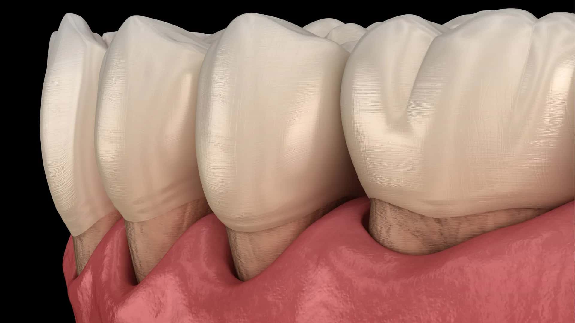
Gum recession around dental implants occurs when the soft tissue surrounding an implant moves away from its original position, potentially exposing the implant crown margin, abutment or implant surface.

- Last updated
- 28 reads
- 15 min read


What Is Gum Recession Around Dental Implants?
Gum recession around dental implants describes a change in the position of the soft tissue margin surrounding an implant-supported tooth. As the tissue moves downward or away from the crown, more of the restoration or underlying implant components may become visible.
The condition is also described as peri-implant mucosal recession or soft tissue dehiscence, particularly when it affects the front-facing surface of an implant.
Recession can create an uneven gum line, expose a darker component beneath the crown or make cleaning more difficult.
However, a visible change in the gum margin does not automatically mean that the implant has lost its connection with the jawbone.
Why Do Gums Recede Around Dental Implants?
Several structural and inflammatory factors can contribute to gum recession around dental implants.
A thin layer of tissue may offer limited coverage over the implant, particularly when the supporting bone on the outer side of the jaw is also thin. Changes in that bone can influence the position of the gum margin.
An implant positioned too close to the outer surface of the jaw may further increase the likelihood of tissue recession.
Inflammation caused by bacterial plaque can damage the surrounding soft tissues and, in more advanced cases, involve the supporting bone.
Crown shape, previous gum disease and changes during healing may also influence the appearance of the implant's gum line.
How Are Gums Around Implants Different From Natural Teeth?
Natural teeth are connected to the surrounding bone through a periodontal ligament, which contributes to their support and tissue organization.
Dental implants integrate directly with the jawbone and do not have the same ligament attachment. The arrangement of connective tissue fibers and blood supply around implants also differs from the structures surrounding natural teeth.
These anatomical differences influence how the soft tissues respond to inflammation, surgery and changes in the supporting bone.
The gum around an implant can remain healthy for many years, but its stability depends on appropriate tissue support, cleaning access and ongoing monitoring.
Recession around an implant therefore requires assessment according to implant-specific anatomy rather than assumptions based entirely on natural teeth.
Can Thin Gum Tissue Increase the Risk of Recession?
Thin peri-implant tissue is an important risk factor for visible recession.
When the tissue covering the front of an implant is delicate, even a relatively small change in its position may expose the underlying restoration or create a gray appearance.
Thin tissue can also make the contour of the implant components more apparent, particularly in the upper front teeth.
Research has associated a thin soft tissue phenotype with a greater likelihood of recession around implants.
However, tissue thickness is not the only consideration. The position of the implant, the amount of supporting bone and the shape of the crown also contribute to long-term stability.
Increasing tissue thickness through grafting may be considered in selected situations.
How Does Implant Position Affect the Gum Line?
The three-dimensional position of a dental implant strongly influences how the surrounding gum tissue is supported.
An implant placed too far toward the lips or cheeks may have limited bone covering its outer surface. This can increase the risk of tissue thinning and recession.
The depth and angle of placement also affect how the final crown emerges through the gum.
Appropriate positioning allows space for supporting bone, soft tissue and a restoration that can be cleaned effectively.
The guide to dental implants, their placement and the healing process explains why implant position and jawbone support matter before the final tooth is attached.
Once an implant has integrated with bone, correcting significant positional problems becomes more complex.
Can Implant Crown Design Cause Gum Recession?
The shape of an implant-supported crown can influence the pressure applied to nearby soft tissues and the ease of daily cleaning.
A restoration with an excessively bulky emergence profile may crowd the gum, create areas where plaque collects or make the implant difficult to clean.
Deep crown margins and inaccessible surfaces can also complicate professional maintenance.
When a crown is cement-retained, residual cement beneath the gum may contribute to inflammation if it is not adequately removed.
However, an unattractive gum line does not always mean the crown was incorrectly designed.
Crown contours must be assessed together with implant position, tissue thickness and bone support before deciding whether prosthetic modification is likely to help.
What Are the Signs of Gum Recession Around Implants?
Gum recession may first become apparent through subtle changes in the appearance of an implant-supported tooth, although additional symptoms can develop when inflammation is present.
-
A longer-looking implant tooth: The crown may appear taller than neighboring teeth because the gum margin has moved away from its previous position.
-
Visible implant components: A dark abutment, metallic edge or portion of the implant surface may become noticeable near the gum line.
-
Uneven gum contours: The tissue around one implant may sit noticeably higher or lower than the gums surrounding adjacent natural teeth.
-
Bleeding or irritation: Redness, bleeding during cleaning and persistent tenderness can accompany recession when peri-implant inflammation is also present.
Is Gum Recession Normal After Dental Implant Surgery?
Some changes in gum position can occur during the initial healing period as surgical swelling resolves and the surrounding tissues mature.
A gum margin that appears slightly different several weeks after surgery does not necessarily indicate an active disease process.
However, noticeable or progressive exposure of the implant surface should not automatically be regarded as normal healing.
Significant tissue loss may reflect inadequate outer bone support, thin gum tissue, surgical changes or an unfavorable implant position.
The distinction becomes clearer when the current gum margin is compared with earlier examinations and photographs.
A healthy healing process generally progresses toward tissue stability rather than continued exposure of deeper implant components.
Can Gum Recession Develop Years After Implant Placement?
Gum recession can develop long after a dental implant has successfully integrated with the jawbone.
The surrounding tissues remain biologically active and can change because of inflammation, bone remodeling and alterations in oral health.
A crown that was originally well covered by gum tissue may become more exposed over time.
Previous periodontal disease, inadequate plaque control and smoking may increase the likelihood of complications affecting the supporting tissues.
Changes can also become more noticeable as neighboring natural teeth and gums change with age.
An implant that has functioned comfortably for several years should therefore continue receiving routine assessments rather than being regarded as permanently free from complications.
Is Implant Gum Recession the Same as Peri-Implantitis?
Gum recession and peri-implantitis are related but distinct conditions.
Recession describes a change in the position of the gum margin. It may occur because of thin tissue or anatomical limitations even when there is no active destructive infection.
Peri-implantitis is an inflammatory disease involving the tissues around an implant and progressive loss of supporting bone beyond expected initial remodeling.
Both conditions can occur together, particularly when inflammation and bone loss contribute to soft tissue changes.
The guide to peri-implantitis, its symptoms and treatment options explains how inflammatory bone loss differs from other changes around dental implants.
Determining whether bone loss is present is essential because gum grafting alone cannot resolve an untreated peri-implant infection.
What Is the Difference Between Peri-Implant Mucositis and Peri-Implantitis?
Peri-implant mucositis involves inflammation of the soft tissue surrounding an implant without progressive loss of supporting bone beyond initial healing changes.
It is commonly associated with bacterial plaque and may cause bleeding during gentle examination, redness or swelling.
Peri-implantitis involves inflammation accompanied by progressive supporting-bone loss. Increased probing depths, bleeding, discharge and changes in the gum margin may also be present.
The distinction matters because mucositis can often improve when plaque-related inflammation is adequately controlled.
Peri-implantitis usually requires a more extensive treatment approach focused on controlling infection and limiting further bone destruction.
Visible recession alone cannot reliably distinguish between these conditions.
Can Gum Recession Expose the Implant Metal or Threads?
As the gum margin recedes, portions of the implant abutment or underlying implant surface may become visible.
In some cases, the exposed material appears as a dark or metallic edge near the crown. More extensive recession, particularly when associated with bone loss, may reveal part of the implant's threaded surface.
However, gray discoloration does not always indicate actual metal exposure.
Very thin gum tissue can allow the color of an underlying component to show through while the surface remains covered.
Direct examination helps distinguish tissue translucency from genuine exposure.
Visible implant threads deserve assessment because they may create cleaning challenges and can indicate a loss of supporting tissue.
Why Is Recession Around Front Tooth Implants More Noticeable?
Implants replacing upper front teeth are particularly sensitive to changes in gum position because the surrounding tissues are easily visible during smiling and speaking.
Even a small difference between the gum margin around an implant and the neighboring natural teeth can affect smile symmetry.
Thin tissue may reveal darker underlying components, while recession can make the implant crown look longer than adjacent teeth.
The curved gum contour and small triangular spaces between teeth also influence the final appearance.
Correcting these changes can be challenging because the cosmetic result depends on both the soft tissues and the position of the implant-supported restoration.
Complete symmetry is not always achievable through grafting alone.
Does Receding Gum Tissue Mean an Implant Is Failing?
Gum recession does not automatically indicate implant failure.
An implant can remain firmly integrated with the jawbone even when the surrounding soft tissue has moved away from its original position.
However, recession should not be ignored because it may coexist with inflammation or changes in bone support.
The implant's stability, surrounding probing measurements and radiographic bone levels help establish whether the problem is primarily cosmetic or involves more serious tissue damage.
A crown or abutment can also become loose independently of the implant body.
The appearance of the gum line therefore provides important information, but it cannot determine implant survival on its own.
How Is Gum Recession Around an Implant Diagnosed?
Assessment begins with examining the gum margin, tissue color, crown shape and visible implant components.
Gentle periodontal probing can measure the depth of the tissue around the implant and identify bleeding or discharge associated with inflammation.
Comparing current measurements with previous records helps reveal whether the gum margin or probing depth has changed.
The amount of firm, keratinized tissue around the implant may also be assessed because limited tissue can make comfortable brushing more difficult.
A complete evaluation includes oral hygiene, implant stability and the design of the restoration.
These findings help distinguish a localized cosmetic defect from a condition affecting the implant's supporting structures.
When Are Dental X-Rays or Additional Imaging Needed?
Dental radiographs help evaluate the bone surrounding an implant and identify changes that cannot be seen through the gum tissue.
Previous images provide a valuable reference for distinguishing expected early bone remodeling from later progressive bone loss.
When bleeding, increased probing depths or discharge raise concern for peri-implantitis, updated radiographs may be necessary.
Standard dental X-rays have limitations, particularly when assessing the thin outer bone wall surrounding an implant.
Three-dimensional imaging may be considered in selected complex cases when additional anatomical detail would affect treatment decisions.
However, imaging should have a clear clinical purpose rather than being used routinely whenever a minor gum contour difference is noticed.
Can Receding Gums Around Implants Grow Back Naturally?
Once gum tissue has moved away from an implant and a stable recession defect has developed, it generally does not return to its original position simply through brushing or home remedies.
Improving oral hygiene may resolve swelling and inflammation, but controlling these symptoms does not automatically replace lost tissue.
In fact, gums can initially appear to recede further as inflamed, swollen tissue returns to a healthier contour.
Where the underlying bone or soft tissue volume is insufficient, rebuilding coverage may require a surgical procedure.
However, complete coverage of exposed implant components is not guaranteed, even after grafting.
The possibility of improvement depends on the defect, implant position and remaining tissue support.
How Is Mild Implant Gum Recession Treated?
Mild recession does not always require immediate surgery, particularly when the implant is stable, the tissues are healthy and the defect is not progressing.
The first priority is identifying whether plaque accumulation, excessive crown contours or another local factor contributes to the condition.
Professional cleaning and suitable home-care techniques can help control inflammation.
If the crown is difficult to clean or places unnecessary pressure on the surrounding tissue, modification may be considered.
Regular measurements allow changes in the gum margin and bone support to be monitored.
However, nonsurgical care primarily aims to stabilize the environment. It should not be presented as a method that predictably restores tissue already lost through recession.
Can a Connective Tissue Graft Cover an Exposed Implant?
Connective tissue grafting is an established surgical approach for increasing soft tissue thickness around selected dental implants.
The procedure often uses connective tissue obtained from the palate. In certain recession defects, the graft can be combined with a coronally advanced flap, which moves the existing gum tissue toward the crown.
This approach may improve tissue thickness and achieve partial or substantial coverage of the exposed area.
However, implant recession defects are not identical to gum recession around natural teeth.
An implant positioned too far toward the lip, extensive tissue loss or unfavorable crown contours may limit the amount of coverage achievable.
Clinical evidence supports selected techniques, but complete and lasting coverage cannot be promised.
What Are the Main Treatment Options for Implant Gum Recession?
Treatment focuses on the specific cause of recession, the health of the implant and the amount of supporting tissue that remains.
-
Inflammation control: Professional plaque removal and improved cleaning access help address gum irritation and reduce bacterial accumulation.
-
Crown or abutment modification: Reshaping or replacing a restoration may improve the emergence profile, cleaning access and pressure on the surrounding tissues.
-
Soft tissue grafting: Connective tissue grafts, selected tissue substitutes or flap procedures may increase tissue thickness and improve coverage in suitable defects.
-
Treatment of supporting-bone disease: When peri-implantitis is present, infection and bone loss must be addressed; advanced cases may require surgery or, when the implant cannot be maintained, removal.
Can Replacing the Implant Crown Improve the Gum Line?
Replacing or modifying the implant crown can sometimes improve the appearance of the surrounding tissue, particularly when the restoration has an unsuitable emergence profile.
A bulky crown may make the gum difficult to clean or create excessive pressure as it passes through the soft tissue.
A more appropriate contour may improve access for brushing and allow the tissues to remain healthier.
In selected cases, crown modification can also support surgical recession treatment by creating a more favorable environment for tissue positioning.
However, changing the crown cannot relocate an implant that has integrated in an unfavorable position or recreate missing jawbone.
Prosthetic correction is therefore most effective when the restoration itself contributes meaningfully to the problem.
What Happens When Gum Recession Occurs With Bone Loss?
Gum recession accompanied by progressive bone loss raises concern for disease affecting the implant's supporting structures.
When peri-implantitis is diagnosed, treatment must address the bacterial biofilm and inflammatory process rather than focusing only on the visible gum margin.
Professional implant-surface cleaning and other nonsurgical measures may form the initial approach.
More advanced disease can require surgical access, implant-surface decontamination and, in selected defects, reconstructive procedures.
Not all bone defects are suitable for regeneration, and soft tissue coverage cannot compensate for uncontrolled infection.
The central objective is to stop further destruction and preserve a stable, maintainable implant whenever possible.
How Long Does Recovery Take After Implant Gum Grafting?
Recovery after an implant gum graft involves healing at both the treated implant area and, when tissue is taken from the palate, the donor site.
Tenderness, swelling and minor bleeding may occur during the first several days.
Initial soft tissue healing generally progresses over approximately one to two weeks, although graft integration and tissue maturation continue much longer.
The final gum contour may develop gradually over several months as swelling resolves and the tissues remodel.
Temporary restrictions on brushing directly over the graft, vigorous rinsing and chewing pressure may be necessary.
A graft that looks healthy after the first week still requires careful protection while its long-term stability develops.
What Are the Risks and Limitations of Gum Recession Treatment?
Surgical correction can improve selected implant recession defects, but the procedure involves risks and does not guarantee complete tissue coverage.
Pain, swelling, bleeding and infection may occur after soft tissue grafting. Harvesting tissue from the palate can also cause temporary discomfort at a second surgical site.
Graft shrinkage, incomplete coverage and recurrent recession are possible outcomes.
An implant positioned too far toward the outer surface of the jaw may remain difficult to cover adequately, even when additional tissue is introduced.
When significant bone loss or active peri-implantitis exists, cosmetic grafting alone cannot resolve the underlying problem.
Long-term success therefore depends on controlling contributing factors as well as healing the surgical site.
How Can Gum Recession Around Dental Implants Be Prevented?
Maintaining stable gums around implants requires attention to both the original treatment design and the long-term health of the surrounding tissues.
-
Maintain effective plaque control: Brush gently but thoroughly, use appropriate interdental cleaning aids and arrange professional implant maintenance.
-
Monitor changes in the gum margin: Regular assessments of bleeding, probing depths and soft tissue levels help identify problems before substantial tissue damage develops.
-
Manage preventable risk factors: Avoid smoking, address existing periodontal disease and maintain appropriate control of health conditions that affect oral tissue healing.
-
Preserve bone and soft tissue support: Suitable implant positioning, adequate tissue thickness and a crown design that permits effective cleaning help reduce the likelihood of future recession.
Which Symptoms Require Prompt Dental Attention?
A newly exposed implant component or a rapidly changing gum margin should be assessed, particularly when the change occurs alongside bleeding or tenderness.
Persistent redness, increasing swelling, discharge and an unpleasant taste may indicate peri-implant inflammation or infection.
Pain that becomes progressively more severe, noticeable implant movement or difficulty chewing also warrants prompt examination.
Rapidly expanding facial swelling, fever accompanied by significant infection symptoms, or difficulty swallowing or breathing requires urgent medical attention.
However, peri-implant disease may progress without severe pain.
For that reason, waiting until an implant becomes uncomfortable is not a reliable way to protect the surrounding bone and gum tissue.
Can Long-Term Implant Health Be Maintained After Gum Recession?
Long-term implant health can often be maintained after gum recession when the underlying cause is recognized and further tissue damage is controlled.
A stable implant with limited recession and healthy surrounding tissues may remain functional without extensive corrective surgery.
Where crown design, inadequate tissue thickness or an inflammatory condition contributes to recession, targeted treatment may improve the environment around the implant.
However, restoring a perfectly symmetrical gum line is not always possible, particularly when the implant position or supporting bone limits treatment.
Ongoing care remains essential even after successful grafting or control of peri-implant inflammation.
The priority should be preserving implant stability, supporting healthy tissues and maintaining effective cleaning access. A natural gum contour is valuable, but the most important long-term result is an implant that remains functional, comfortable and free from progressive inflammatory damage.
- Last updated
- Language
- English
How did you find this article?
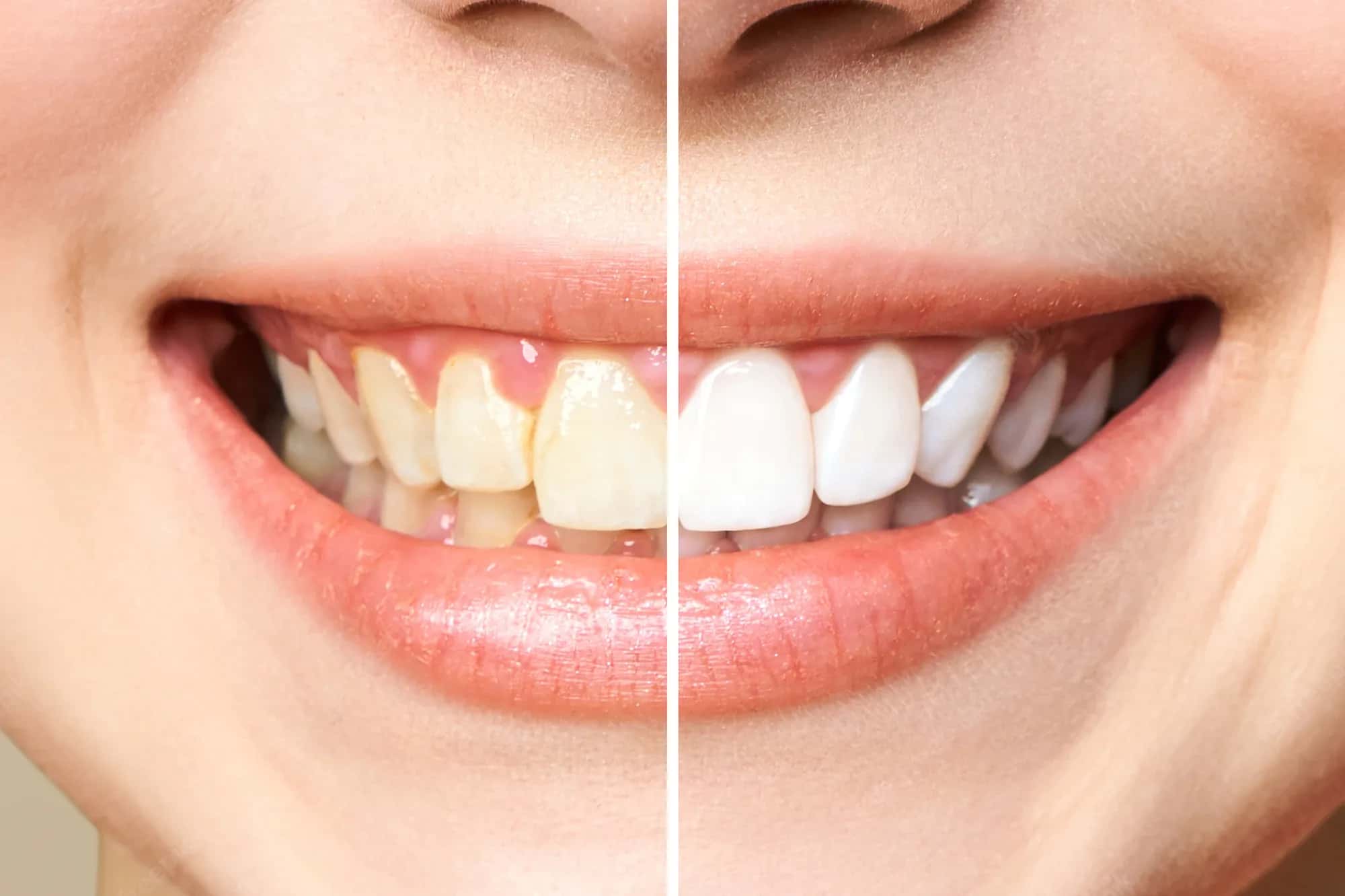
Teeth whitening costs around the world vary substantially, with professional treatment generally costing less in countries such as Turkey, Mexico and India than in the United States or United Kingdom.
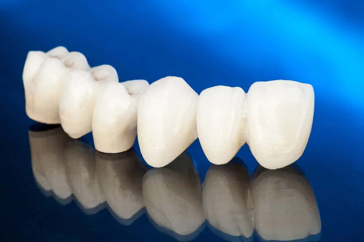
Zirconium crown costs around the world vary significantly, from approximately €180–€400 per tooth in Turkey to $1,000–$2,500 in the United States, based on published 2026 private-treatment prices.
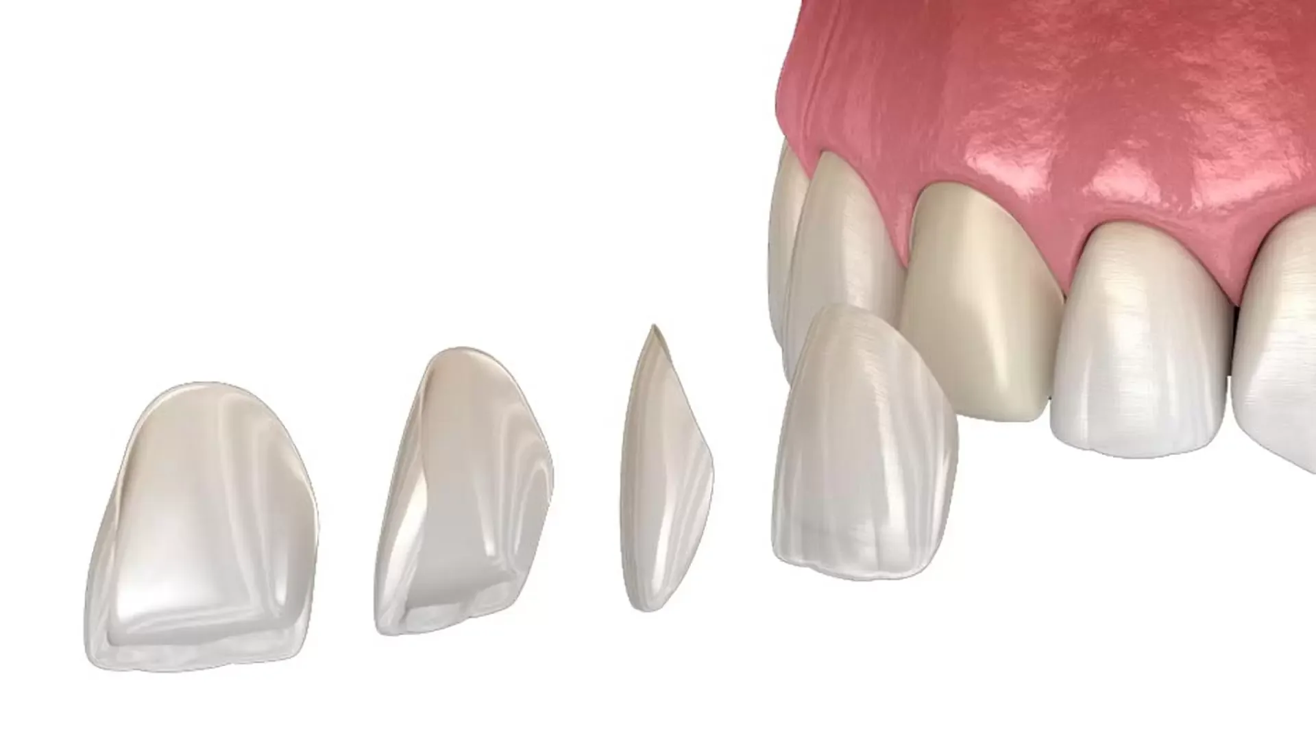
E-Max costs around the world differ significantly, with ceramic veneers and crowns available at lower published prices in countries such as Turkey, Mexico and Hungary than in the United States or United Kingdom.
Subscribe to the Medifinder newsletter and never miss our latest content.