










Dental Implants With Bone Loss
Jawbone loss can make dental implant treatment more complex, but bone grafting, sinus lifts and specialized implant designs give many patients a path to a stable replacement tooth.

- Last updated
- 19 reads
- 10 min read


Can You Get Dental Implants If You Have Bone Loss?
Many people with jawbone loss can still receive dental implants, although they often need additional procedures or a different implant approach to create enough support. An implant relies on direct contact with healthy bone, a process called osseointegration, so the bone around it must be tall, wide and dense enough to hold it firmly while it heals and later while it carries chewing forces.
When bone has thinned, the dentist or oral surgeon has two broad options. The first is to rebuild the missing bone with grafting, which adds volume to the jaw before or during implant placement. The second is to choose implants and placement techniques that make use of the bone that remains, such as shorter implants, angled implants or implants anchored in denser areas of the skull.
The right choice depends on how much bone has been lost, where the loss is located and the patient's general health. A detailed examination and 3D imaging usually come first, since they show exactly what the jaw can support.
Why Does the Jawbone Shrink After Tooth Loss?
The jawbone shrinks after tooth loss because the bone that once held the tooth no longer receives the stimulation it needs to maintain itself. Every time you bite and chew, your teeth pass forces through their roots into the surrounding bone, and this pressure signals the body to keep that bone in place. Once a tooth is gone, the area around the empty socket gradually loses volume in a process called resorption.
Resorption often moves fastest in the first months after an extraction and then continues more slowly over the years. The ridge of bone usually loses width first and then height, which can leave a narrow, flattened edge where a full ridge once stood.
Other factors speed up or add to this process. Advanced gum disease, known as periodontitis, destroys the bone that supports the teeth even before they fall out. Long-term use of removable dentures can press on the ridge and contribute to further shrinkage. Infections, cysts, facial trauma and difficult extractions can also remove bone, and in the upper back jaw, the sinus cavity can expand downward into the space left by missing molars.
What Are the Signs of Bone Loss in the Jaw?
Jawbone loss often develops quietly, yet several changes in the mouth and face can point to it before any imaging confirms it:
- Dentures that become loose or uncomfortable over time, since the ridge beneath them changes shape and no longer matches the fitting surface.
- Teeth that shift, tilt or drift into the space left by a missing tooth, which changes the bite and can affect the bone around neighboring teeth.
- Receding or shrinking gums around missing teeth, often with a visible dip or narrowing in the gum line where the bone has thinned.
- Changes in facial appearance, including a sunken look around the mouth, thinner lips or a shorter distance between the nose and chin when many teeth are missing.
- Loose teeth or deep gum pockets in people with gum disease, which suggest that bone supporting the remaining teeth is breaking down.
How Do Dentists Measure Bone Before Implant Placement?
Dentists measure bone before implant placement mainly with cone beam computed tomography, a 3D scan that shows the height, width and density of the jaw along with nearby anatomical structures. Standard dental X-rays provide a flat, two-dimensional view, which can hide narrow ridges or make bone look more plentiful than it really is.
The 3D scan lets the clinician see the position of the nerve in the lower jaw, the floor of the maxillary sinus in the upper jaw and any defects or thin areas in the ridge. These details decide how long and wide the implant can be, which angle it should take and whether grafting is necessary to place it safely.
Many implant teams also use the scan with digital planning software, which helps them position implants virtually before surgery. In some cases, they produce a surgical guide from this plan, a custom template that directs the drill to the planned position and angle during the procedure.
How Does Bone Grafting Prepare the Jaw for Implants?
Bone grafting prepares the jaw for implants by adding material to areas where bone is missing, which gives the body a framework to grow new bone into over time. As healing progresses, the graft gradually becomes part of the jaw and provides a stable base that can hold an implant.
A surgeon may place a graft before the implant and allow it to heal for several months, or place the graft and implant in the same appointment when enough bone already exists to stabilize the implant from the start. The amount and location of the bone loss guide this decision.
Socket Preservation and Ridge Augmentation
Socket preservation takes place at the time of tooth extraction, when the surgeon fills the empty socket with graft material to reduce the shrinkage that usually follows. This step can make later implant placement simpler by keeping more of the original ridge.
Ridge augmentation rebuilds a ridge that has already lost width or height. The surgeon places graft material along the deficient area and often covers it with a protective membrane, a technique called guided bone regeneration, which keeps soft tissue out and allows bone to fill the space. For larger defects, the surgeon may use a block of bone fixed to the jaw with small screws.
Sinus Lift for the Upper Back Jaw
A sinus lift adds bone to the upper back jaw, where the maxillary sinus often sits close to the ridge after molars are lost. The surgeon gently lifts the thin membrane that lines the floor of the sinus and places graft material beneath it, which increases the bone height available for implants.
When only a small amount of extra height is needed, the surgeon can approach the sinus through the implant site itself. For larger lifts, they create a small window in the side wall of the upper jaw to reach the sinus floor more directly. Both approaches aim to create enough bone to hold implants securely in an area that would otherwise be too thin.
Which Bone Graft Materials Are Used?
Surgeons choose from several types of graft material, and each one offers a different balance of biological activity, availability and comfort for the patient:
- Autografts: Bone taken from the patient's own body, often from the chin, the back of the jaw or occasionally the hip, which carries living cells and integrates well but requires a second surgical site.
- Allografts: Processed bone from human donors, which tissue banks sterilize and prepare carefully, and which avoids the need to harvest the patient's own bone.
- Xenografts: Bone mineral from animal sources, most commonly bovine, which serves as a slowly resorbing scaffold that helps maintain volume while new bone forms.
- Synthetic grafts: Laboratory-made materials such as calcium phosphate compounds, which provide a framework for bone growth without any biological donor.
- Growth factor additions: Concentrates prepared from the patient's own blood, such as platelet-rich fibrin, which some surgeons add to grafts with the aim of supporting soft tissue and bone healing.
Are There Implant Options That Avoid Bone Grafting?
Several implant approaches can reduce or avoid the need for bone grafting by working with the bone that is still present. These options do not suit every situation, but they give patients with significant bone loss more choices and sometimes a shorter overall treatment time.
Short and Narrow Implants
Short implants have a reduced length, which allows placement in areas where limited bone height sits above the nerve in the lower jaw or below the sinus in the upper jaw. Narrow implants, sometimes called mini or reduced-diameter implants, fit into thinner ridges where a standard implant would leave too little bone around it. Clinicians select these designs carefully, since the forces they can withstand depend on their size, the bite and the restoration they support.
Angled Implants and Full-Arch Solutions
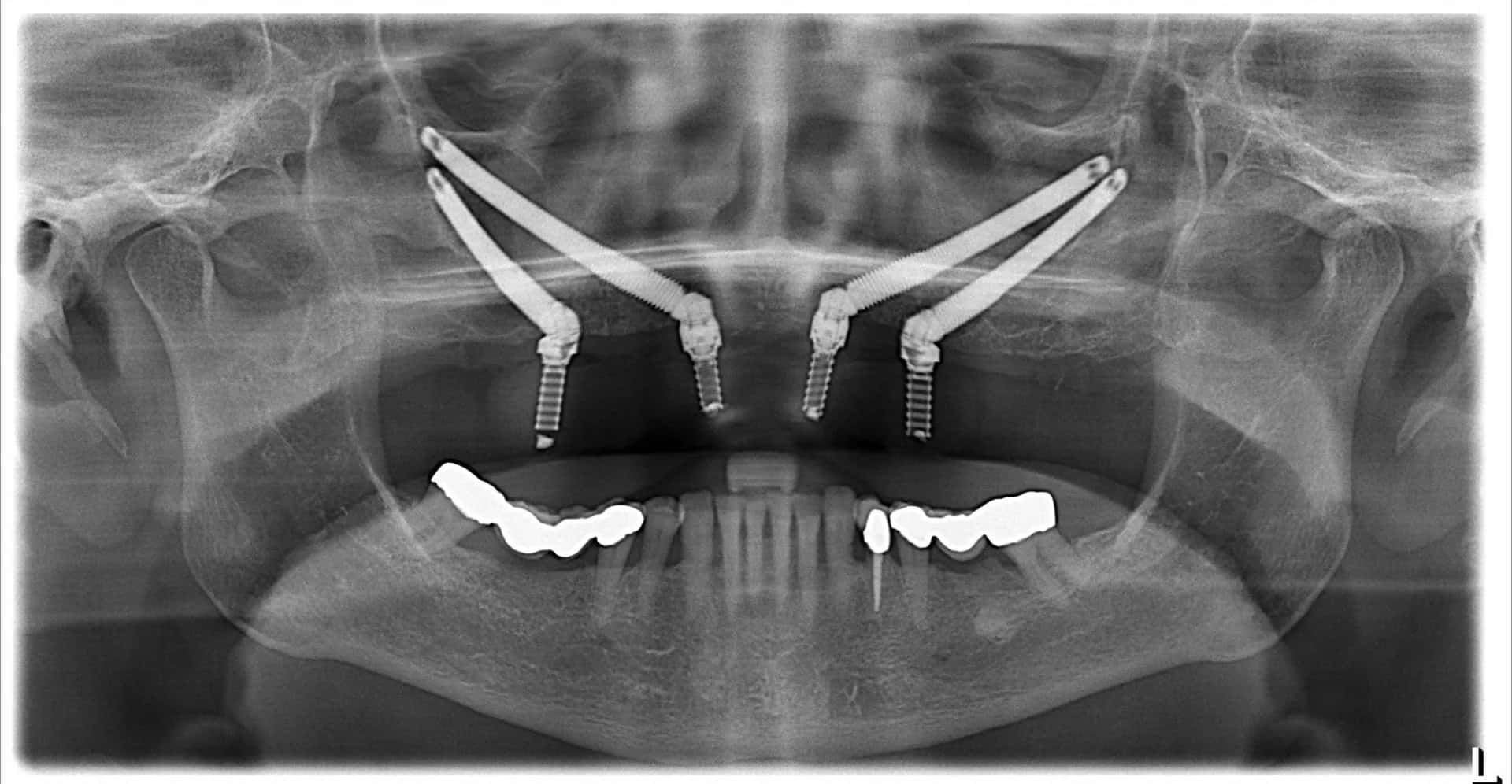
For patients missing all or most of their teeth in one jaw, full-arch solutions can support a fixed bridge on four to six implants. In these designs, the surgeon often tilts the back implants so they avoid the sinus or the nerve and anchor in denser bone toward the front of the jaw. This angled placement makes better use of existing bone and may allow a fixed temporary bridge soon after surgery when the implants achieve enough initial stability.
Zygomatic Implants for Severe Upper Jaw Bone Loss
Zygomatic implants are long implants that pass through the upper jaw and anchor in the cheekbone, known as the zygoma, which keeps dense bone even when the upper jaw has resorbed severely. They offer an option for patients who would otherwise need extensive grafting or who have experienced failed grafts. Because they involve complex anatomy close to the sinus and the eye socket, specialists with specific training in this technique usually perform them.
How Long Does Treatment Take When Bone Loss Is Present?
Treatment usually takes longer when bone loss is present, since grafted bone needs time to mature before it can reliably support an implant. A ridge augmentation or sinus lift commonly requires several months of healing before implant placement, and the implant itself then needs further time to integrate with the bone before the final crown or bridge is attached.
When the surgeon can place the graft and implant together, the overall timeline becomes shorter because both heal at the same time. Approaches that avoid grafting altogether, such as angled full-arch implants or zygomatic implants, can shorten treatment further and sometimes allow a temporary fixed restoration soon after surgery.
During healing, patients usually wear a temporary tooth, bridge or denture to maintain appearance and function. The final restoration follows once the clinician confirms that the implant is stable and the surrounding tissues have healed well.
What Risks Should You Consider?
Implant treatment with bone grafting carries the usual risks of oral surgery along with a few that relate specifically to grafts and sinus procedures. Swelling, bruising, discomfort and minor bleeding are common in the first days, and most people manage them with prescribed medication and careful aftercare.
Less common complications include infection, exposure of the graft or membrane through the gum, partial loss of the graft and insufficient bone formation, which may require a repeat procedure. A sinus lift can tear the sinus membrane, and the surgeon may repair it during the same appointment or stop and allow it to heal before trying again. In the lower jaw, implants placed close to the nerve carry a risk of numbness or altered sensation in the lip and chin.
General health also shapes the risk profile. Smoking slows healing and raises the chance of graft and implant problems. Poorly controlled diabetes, certain bone-related medications and a history of radiation to the head and neck all need careful assessment, so sharing your full medical history at the first consultation matters.
What Should You Discuss Before Starting Treatment?
A thorough conversation before treatment helps you understand the plan, the timeline and the alternatives available for your level of bone loss:
- What the 3D scan shows, including how much bone is available, where the deficiencies are and how close the planned implants will sit to the nerve or sinus.
- Whether grafting is necessary, which type of graft the surgeon recommends and whether the graft and implant can be placed in the same appointment.
- Graft-free alternatives, such as short, narrow, angled or zygomatic implants, and how they compare with grafting for your situation.
- The expected timeline, from extraction or grafting through healing to the final restoration, along with the type of temporary teeth you will wear in between.
- Your health and lifestyle factors, including smoking, medications and medical conditions, and the steps that can lower your risk of complications.
Finding an Implant Specialist for Complex Cases
Bone loss often turns a routine implant into a more advanced surgical case, so the experience of the treating team matters. Look for a qualified oral surgeon, periodontist or implant dentist who uses 3D imaging, explains both grafting and graft-free options clearly and can show cases with a similar level of bone loss.
MediFinder lets you browse dental clinics and doctor profiles, compare the implant and bone grafting services they list and send an appointment request when you find a team that suits your needs. A careful assessment of your jawbone is the first step toward a treatment plan that gives your new teeth a stable foundation.
- Last updated
- Language
- English
How did you find this article?
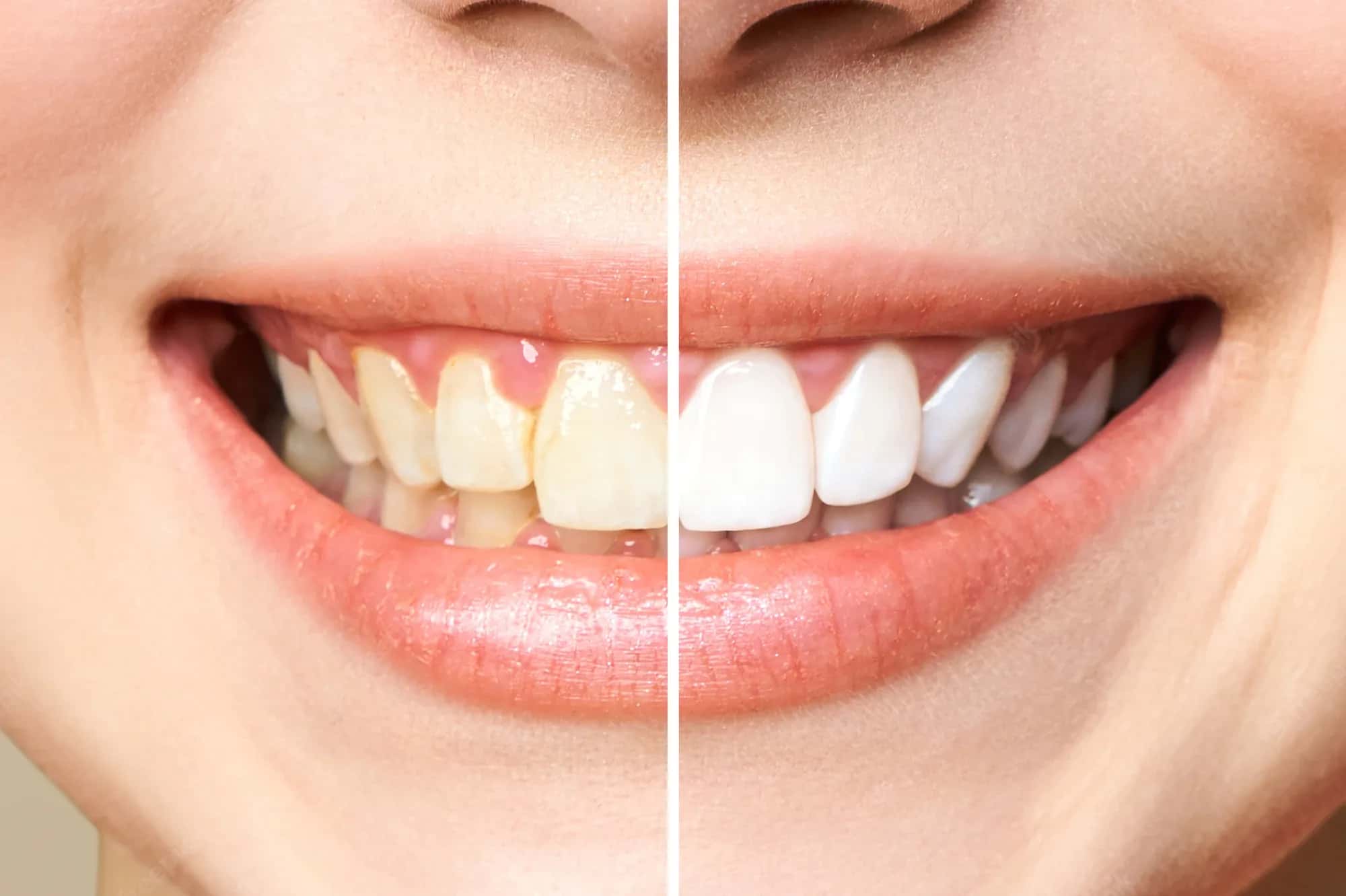
Teeth whitening costs around the world vary substantially, with professional treatment generally costing less in countries such as Turkey, Mexico and India than in the United States or United Kingdom.
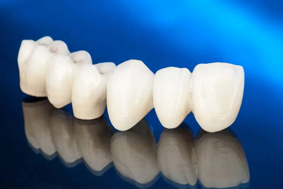
Zirconium crown costs around the world vary significantly, from approximately €180–€400 per tooth in Turkey to $1,000–$2,500 in the United States, based on published 2026 private-treatment prices.
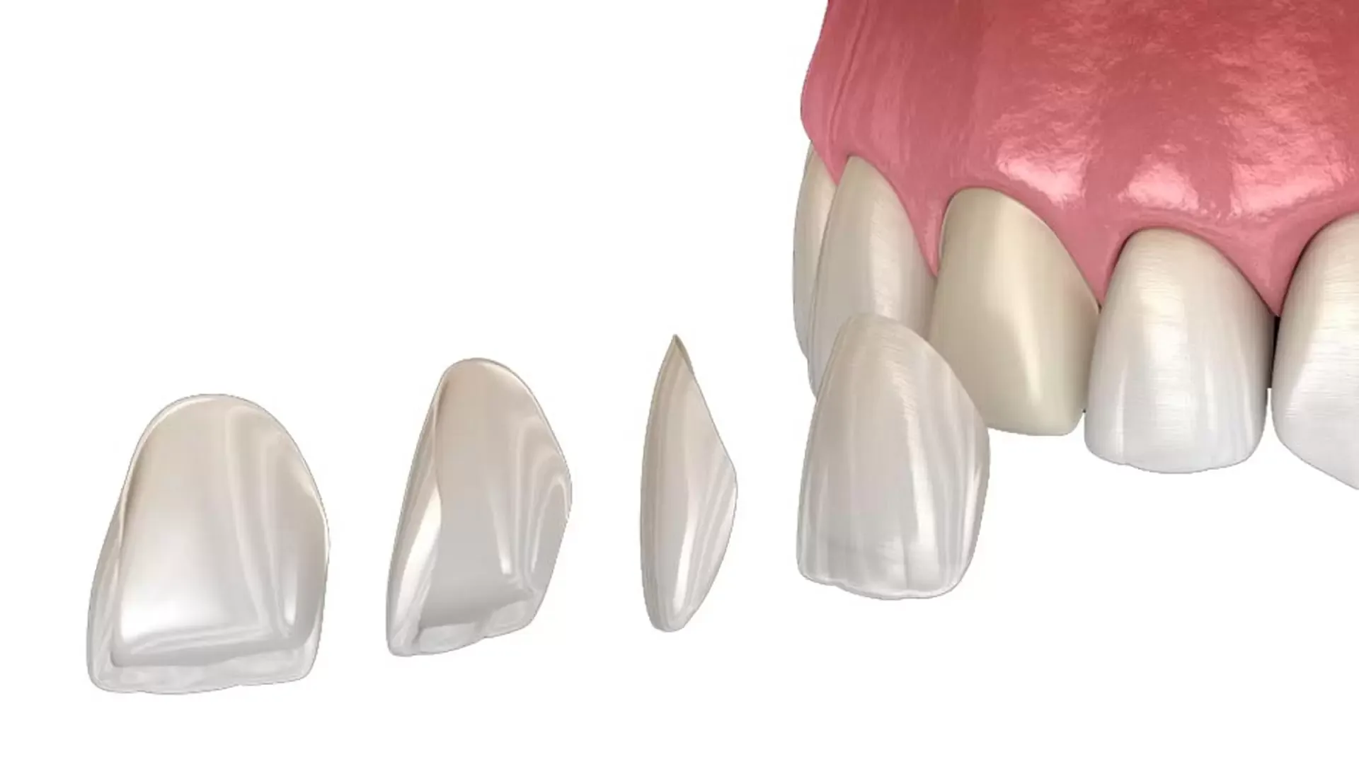
E-Max costs around the world differ significantly, with ceramic veneers and crowns available at lower published prices in countries such as Turkey, Mexico and Hungary than in the United States or United Kingdom.
Subscribe to the Medifinder newsletter and never miss our latest content.